AORTIC VALVE
TAVR
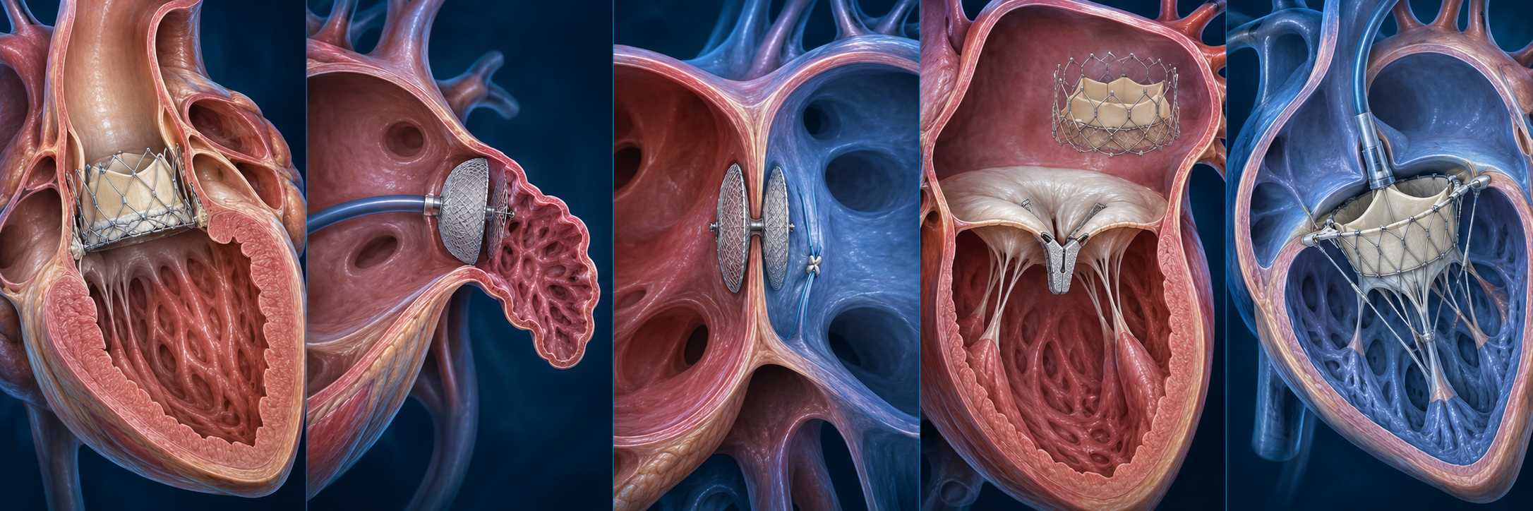
A bioprosthetic valve frame positioned within the native aortic annulus.
Physician-informed research and development focused on structural heart valve technologies, catheter-based therapies, and elegant solutions for complex anatomy.
Explore the target anatomy and the general form of catheter-based technologies used across contemporary structural heart care.
A bioprosthetic valve frame positioned within the native aortic annulus.
A conformable occlusion implant seated at the appendage ostium.
Disc-based occlusion and tissue-preserving suture closure concepts.
Leaflet approximation and transcatheter replacement concepts for mitral disease.
Catheter-based repair and replacement concepts designed for right-heart anatomy.
Educational illustrations are generic and are not depictions of any specific manufacturer’s product. Device selection and treatment decisions require evaluation by a qualified heart team.
Our focus spans valve replacement, repair, occlusion, and closure systems—grounded in anatomy, procedural reality, and meaningful clinical need.
Concepts for valve performance, anchoring, sealing, commissural alignment, coronary access, delivery systems, and lifetime management.
Approaches to adaptable fixation, complete sealing, lower device footprint, procedural simplicity, and anatomy-responsive closure.
Transcatheter and suture-based concepts for ASD and PFO closure with attention to tissue preservation and future access.
Replacement and repair concepts addressing annular complexity, LVOT interaction, sealing, anchoring, and deliverability.
Systems designed around large, dynamic anatomy, variable leaflet geometry, right-heart physiology, and venous access.
A disciplined pathway links clinical insight to engineering development while keeping usability, anatomy, and safety at the center.
Identify a consequential clinical or procedural limitation worth solving.
Translate imaging, biomechanics, and procedural constraints into requirements.
Develop concepts, assess failure modes, and refine through structured testing.
Prepare for verification, preclinical evaluation, regulatory strategy, and translation.
Delivery, visualization, deployment, repositioning, retrieval, and workflow should be designed as one integrated clinical experience—not addressed after the device is built.
Structural Heart Innovations brings a procedural lens to early-stage device strategy, concept definition, and development planning.
Procedural pain points, patient selection gaps, anatomical limitations, workflow friction, and complication-driven opportunities.
Physician-led product requirements, delivery concepts, deployment sequences, and usability priorities.
CT, echocardiographic, fluoroscopic, and access considerations integrated into device architecture.
Bench-test logic, preclinical questions, risk framing, clinical endpoints, and development milestones.
Engagement with engineers, inventors, research partners, manufacturers, and development teams.
The complete advanced module now includes 16 chapters covering guideline-based selection, quantitative CT and TEE, device-specific mechanics, geometry-driven transseptal access, implant troubleshooting, rescue algorithms, antithrombotic pathways, surveillance, trial-level outcomes, and linked primary literature.
In nonvalvular atrial fibrillation, thrombus frequently originates in the left atrial appendage. Percutaneous LAA closure mechanically excludes this trabeculated, low-flow chamber from the systemic circulation. The therapeutic trade is deliberate: accept an up-front invasive procedure and a period of device healing to reduce long-term dependence on oral anticoagulation.
CHA₂DS₂-VASc estimates thromboembolic risk and HAS-BLED helps reveal modifiable bleeding hazards, but neither substitutes for judgment. Document the bleeding phenotype, its reversibility, expected anticoagulant exposure, life expectancy, frailty, competing stroke mechanisms, and the patient’s goals.
| Favors closure | Requires deliberation | Usually defer / avoid |
|---|---|---|
| Prior intracranial hemorrhage; recurrent GI bleeding; major bleeding on appropriate OAC; very high future bleeding risk | Remote corrected bleed; isolated preference; severe CKD; active cancer; very elderly or frail patient | Active LAA thrombus; infection; incompatible anatomy; inability to take required post-implant therapy; very limited life expectancy |
The LAA ostium lies anterolateral to the left superior pulmonary vein and superior to the mitral annulus, with the left circumflex artery and great cardiac vein along its inferior epicardial relationship. Chicken-wing, windsock, cactus, and cauliflower labels are descriptive—not procedural endpoints. What matters is the ostial plane, landing-zone diameter, depth, angulation, lobes, trabeculation, proximal ridge, and relationship to the pulmonary vein and mitral apparatus.
The puncture determines coaxiality, sheath torque, depth, and deployment force. A posterior-inferior puncture is frequently favorable, but the ideal site is anatomy- and device-specific. Use bicaval and short-axis TEE views, confirm tenting, and assess the trajectory before crossing. An anterior puncture can force oblique deployment; an excessively superior puncture can reduce working depth.
Interrogate the LAA near 0°, 45°, 90°, and 135° with deliberate gain and depth optimization. Identify thrombus versus spontaneous echo contrast, measure the ostium and intended landing zone, define maximum diameter and usable depth, evaluate lobes, document baseline pericardial space, and inspect the interatrial septum.
Use ECG-gated cardiac CT with adequate left-atrial opacification; a delayed phase helps distinguish slow flow from thrombus. Reconstruct the ostial plane orthogonal to the LAA axis and measure minimum and maximum diameters, perimeter, area, depth, and morphology. CT often reveals a larger maximal dimension than 2-D TEE because the landing zone is elliptical.
| Question | TEE strength | CT strength |
|---|---|---|
| Exclude thrombus | Real-time flow and spontaneous contrast | Excellent with delayed phase |
| Size landing zone | Dynamic and intraprocedural | True orthogonal 3-D geometry |
| Plan puncture | Direct procedural guidance | Pre-plans height and trajectory |
| Assess seal | Color Doppler | High-resolution position and patency |
| Design | Mechanism | Key consideration |
|---|---|---|
| Single-lobe / plug | Endocardial plug seated in a landing zone | Depth, circumferential engagement, compression, and stable proximal position |
| Lobe-and-disc | Distal lobe anchors while a proximal disc covers the ostium | Disc orientation, mitral/PV clearance, lobe compression, and ostial coverage |
| Complication | Early signal | Immediate response |
|---|---|---|
| Perforation / tamponade | Hypotension, new effusion, chamber collapse, tachycardia | Stop manipulation; reverse when appropriate; urgent drainage, blood, and surgical support |
| Device embolization | Loss of position or visualization in LA, LV, or aorta | Localize and plan snare retrieval versus surgery without worsening obstruction |
| Air / thromboembolism | ST change, neurologic deficit, collapse | 100% oxygen, aspiration when accessible, coronary/cerebral emergency pathway |
| Peridevice leak | Residual color jet or CT patency | Characterize mechanism and size; optimize before release when feasible |
| Device-related thrombus | Mass or hypoattenuated thickening | Anticoagulate when safe and repeat imaging to document resolution |
| Access bleeding | Pain, swelling, falling hemoglobin | Compression, ultrasound, reversal/transfusion or vascular intervention |
The evidence has evolved from warfarin-controlled efficacy trials to contemporary device registries, randomized comparison with DOACs, head-to-head device trials, and ablation-adjacent strategies. Interpret each study by its comparator, device generation, post-implant regimen, operator era, and enrolled bleeding risk.
Established proof of concept versus warfarin, with long-term thromboembolic protection but an early procedural safety penalty from the first-generation era.
Added randomized warfarin-comparator evidence and improved procedural safety while highlighting endpoint and noninferiority nuances.
Directly compared closure with DOAC therapy in high-risk patients and supported noninferiority for a composite of ischemic, bleeding, and procedural outcomes.
Prospective evaluation showing high implant success and favorable seal with the redesigned Watchman FLX platform; nonrandomized.
Randomized Amulet versus Watchman: noninferior safety and effectiveness with superior anatomic closure, balanced against device-specific procedural considerations.
After AF ablation, closure reduced clinically relevant nonprocedural bleeding and was noninferior for death, stroke, or systemic embolism versus anticoagulation.
Antithrombotic therapy must follow current device labeling, the evidence-supported regimen, and the patient’s bleeding and thrombotic risk. Contemporary strategies may use short-term anticoagulation plus aspirin or dual antiplatelet therapy in selected circumstances. No single regimen is universal.
TEE or cardiac CT is commonly performed during early follow-up, with timing determined by labeling, regimen, protocol, and clinical events. New neurologic symptoms, embolism, unexplained dyspnea, chest pain, or bleeding should trigger earlier evaluation.
Educational material for clinicians. It is not a substitute for device-specific training, current instructions for use, institutional protocols, multidisciplinary judgment, or individualized care. Evidence and labeling evolve; verify the current primary source before clinical application. Last educational review: September 2026.
Dr. Raj Marok is a board-certified interventional and structural cardiologist with more than a decade of experience in complex coronary intervention, transcatheter valve therapy, left atrial appendage closure, and congenital structural interventions. A Clovis native, he returned to California’s Central Valley after serving as Medical Director of Structural Heart Disease and Co-Director of a complex high-risk PCI and CTO program in West Michigan.
He previously served as an Associate Professor of Medicine at Michigan State University College of Human Medicine and as core faculty for an interventional cardiology fellowship. His advanced interventional training included structural heart, complex coronary, and vascular intervention at Columbia University Medical Center and NewYork-Presbyterian Hospital.
Dr. Marok’s research and development interests center on left atrial appendage closure, transcatheter valve technologies, imaging-guided intervention, and quality improvement. He has served as a principal investigator and sub-investigator in multicenter clinical trials and participated in the world’s first preclinical implant of the WATCHMAN FLX Pro device as part of its FDA approval program.
His work is driven by a simple premise: device design should begin with anatomy, workflow, and the unmet needs encountered in real patient care.
If you are interested in learning about our research focus, sharing a patient perspective, or receiving future updates, send us a brief message.
We are developing and evaluating new ideas at the intersection of structural heart medicine, device engineering, and procedural innovation.